

The World's Lightest Electromagnetic Shielding Material
August 14, 2020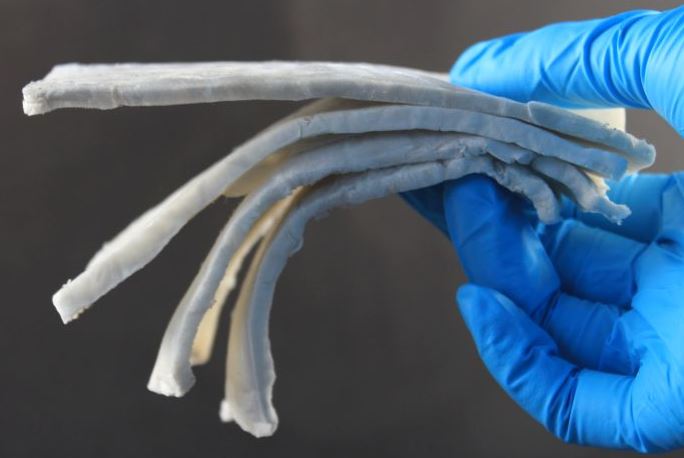 Electric motors and electronic devices generate electromagnetic fields that sometimes have to be shielded in order not to affect neighboring components or the transmission of signals. However, the thin metal sheets or metallized foils that are conventionally used for this purpose are often too heavy or too rigid for many applications. To address this shortcoming, Empa researchers recently succeeded in applying very light aerogels, based on cellulose nanofibers, to microelectronics, which can effectively shield electromagnetic radiation over a wide frequency range. Cellulose fibers are obtained from wood, and due to their chemical structure, enable a wide range of chemical modifications, which are maintained even after being subjected to strong mechanical stress. Combined with two-dimensional nanoplates of titanium carbide, the material has proven to be the lightest electromagnetic shield in the world.
For more details, see here.
Microgripper Captures Single Cells with Magnetic Help
August 02, 2020.JPG) Sampling diseased tissue, which can contain many different types of cells, calls for precise biomedical tools. Current methods for manipulating single cells, like laser microdissection, require complicated setups and can damage cells. Now, researchers led by David H. Gracias at Johns Hopkins University have developed devices that could offer a simpler and less-harmful way to isolate single cells (Nano Lett. 2020, DOI: 10.1021/acs.nanolett.0c01729). Remotely guided by a magnetic field, these so-called microgrippers can wrest single cells from a tissue sample and carry them on demand. Though the tiny grippers require more optimization and testing, the researchers hope one day to use them to perform single-cell biopsies inside a human body.
Sampling diseased tissue, which can contain many different types of cells, calls for precise biomedical tools. Current methods for manipulating single cells, like laser microdissection, require complicated setups and can damage cells. Now, researchers led by David H. Gracias at Johns Hopkins University have developed devices that could offer a simpler and less-harmful way to isolate single cells (Nano Lett. 2020, DOI: 10.1021/acs.nanolett.0c01729). Remotely guided by a magnetic field, these so-called microgrippers can wrest single cells from a tissue sample and carry them on demand. Though the tiny grippers require more optimization and testing, the researchers hope one day to use them to perform single-cell biopsies inside a human body.
For more information, and a movie, check it out here.
Special Issue Planned About Magnetic Cell Separation
June 02, 2020 Our colleague Dr. Marie Frenea-Robin is the guest editor of a special issue of magnetochemistry, titled "Magnetic Cell Separation". Magnetic cell separation has become a key methodology for the isolation of target cell populations from biological suspensions, covering a wide spectrum of applications from diagnosis and therapy in biomedicine to environmental applications or fundamental research in biology. This Special Issue aims to create a forum of discussion to share advances and address current challenges in magnetic cell separation. For more details, check out this flyer.
The deadline for your manuscript submission will be November 30, 2020.
List of Cancelled Magnetic Conferences / Potential Jobs in Magnetism
May 01, 2020 The European Magnetism Society just sent out a newsletter with a list of cancelled magnetism-related meetings. Check it out here:
http://magnetism.eu/news/144/38-news.htm
Also, while the Covid-19 situation is still dire, the curve seems flattened in many countries, and governments are carefully starting to reopen schools etc. This also means that more people are looking for jobs again.
In the magnetism area, EMA also updated their lists of available jobs. Please check it out too:
http://magnetism.eu/5-job-market.htm
And if you are an employer who is looking for a new employee, please advertise at EMA, or also send us an e-mail so we can advertise on magneticmicrosphere.com.
Virtual MPI Meetings Organized by Magnetic Insight
April 20, 2020 In the light of major conference cancellations, the magnetic particle imaging community has decided to maintain our fast progress in molecular imaging and support the trainees that are hard at work producing groundbreaking data.
MPI is inviting researchers in the MPI field to participate in the Magnetic Particle Imaging Rising Stars 2020 Molecular Imaging eSymposia, taking place on April 21st and 22nd at 9 AM PT / 12 PM ET.
If you do some cool MPI research and would like to give a short 10 minute presentation, then please submit an abstract online at this address.
Abstract Deadline: April 3rd, 2020. For any questions, please contact Jeff Gaudet PhD.
Full-Color Magnetic Nanoparticles Based on Holmium-Doped Polymers
April 08, 2020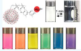 Michinari Kohri et al. just published a very cool paper in ACS Applied Polymer Materials. They demonstrated the production of colorless and full-color magnetic nanoparticles based on holmium (Ho)-doped polymers, which could not be achieved with conventional dark brown iron oxide magnetic nanoparticles. The coordination of Ho, a lanthanide with low colorability and a strong magnetic moment, with a poly(2-acetoacetoxy ethyl methacrylate) brush built on the surface of submicron-sized silica particles allowed for the formation of colorless magnetic nanoparticles. Additionally, bright and full-color magnetic nanoparticles were obtained by mixing different colored magnetic nanoparticles that were prepared by copolymerization of 2-acetoacetoxy ethyl methacrylate and dye monomers. Various colors, including transparency, were demonstrated by means of the present method, which determines the presence or absence of magnetism by Ho doping.
The bright and magnetically controllable colored nanoparticles presented herein may have a significant impact on practical substances and applications, such as ink and biomedical and device applications.
Check out the article here.
200th Anniversary of Hans Christian Oersted
April 07, 2020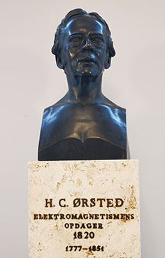 On the occasion of the 200th anniversary of Hans Christian Ørsted's discovery of electromagnetism, a number of events were held during the year, including a series of anniversary lectures, which unfortunately had to be canceled due to the Coronavirus situation. Several of them have been video-recorded and can be viewed on YouTube.
As many of you know, I have a joint professorship at the Department of Pharmacy at the University of Copenhagen. Oersted was a pharmacist, and we can see him every day as a bronze bust (see to the right).
If you interested in his history, I recommend to watch now one of these lectures: HC Ørsted and the Pharmacy History, by Dr. Pharm. Poul R. Kruse, who worked at the Danish School of Pharmacy. With his usual legendary thoroughness and accuracy, Poul Kruse examines HC Ørsted's pharmaceutical background and decisive influence on pharmaceutical (and chemistry) education in Denmark in the 19th century, including the importance for the later development of the Danish pharmaceutical and chemical industry.
You can watch the lecture via this link: https://www.youtube.com/watch?v=oMIBqtR7f5Q
Magnetic Particle Spectroscopy for Detection of Influenza A Virus Subtype H1N1
April 06, 2020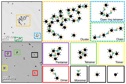 Magnetic nanoparticles (MNPs) with proper surface functionalization have been extensively applied as labels for magnetic immunoassays, carriers for controlled drug/gene delivery, tracers and contrasts for magnetic imaging, etc. Kai Wu and Jian-Ping Wang et al. introduced a new biosensing scheme based on magnetic particle spectroscopy (MPS) and the self-assembly of MNPs to quantitatively detect H1N1 nucleoprotein molecules. MPS monitors the harmonics of oscillating MNPs as a metric for the freedom of rotational process, thus indicating the bound states of MNPs. These harmonics can be readily collected from nanogram quantities of iron oxide nanoparticles within 10 s. The H1N1 nucleoprotein molecule hosts multiple different epitopes that forms binding sites for many IgG polyclonal antibodies. Anchoring IgG polyclonal antibodies onto MNPs triggers the cross-linking between MNPs and H1N1 nucleoprotein molecules, thereby forming MNP self-assemblies. Using MPS and the selfassembly of MNPs, they were able to detect as low as 44 nM (4.4 pmole) H1N1 nucleoprotein. In addition, the morphologies and the hydrodynamic sizes of the MNP self-assemblies are characterized to verify the MPS results. Different MNP self-assembly models such as classical cluster, open ring tetramer, and chain model as well as multimers (from dimer to pentamer) are proposed and form a new biosensing scheme for detecting ultralow concentrations of target biomolecules as a rapid, sensitive, and wash-free magnetic immunoassays. Check out more details in ACS Applied Materials & Interfaces.
For more information, check out our Archives.
September 2017
Search this site with the power of